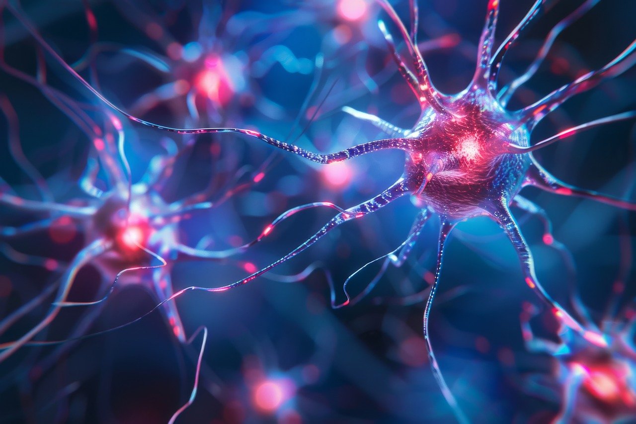
The findings shed new light on the functioning of neurons.Continue reading


Easy Branches allows you to share your guest post within our network in any countries of the world to reach Global customers start sharing your stories today!
Easy Branches
34/17 Moo 3 Chao fah west Road, Phuket, Thailand, PhuketCall: 076 367 766
info@easybranches.comThe joint research of the Columbia University’s Zuckerman Institute in New York, the BrainVisionCenter led by Balázs Rózsa, and the HUN-REN Institute of Experimental Medicine (HUN-REN IEM) has produced a revolutionary result: for the first time

The joint research of the Columbia University’s Zuckerman Institute in New York, the BrainVisionCenter led by Balázs Rózsa, and the HUN-REN Institute of Experimental Medicine (HUN-REN IEM) has produced a revolutionary result: for the first time ever, the birth of memories has been observed in structures 100 times thinner than a human hair. The discovery could open up new perspectives in the treatment of aging and neurological diseases, writes the HUN-REN Hungarian Research Network’s website.
The Hungarian-developed 3D laser scanning microscope is the first to observe the birth of memories in living animals in a fraction of a second in structures 100 times thinner than a human hair. The study was published in the prestigious scientific journal Nature.
Recall of memories is based on changes in the strength of connections between brain cells, known as synapses. Although this has been known for almost 50 years, scientists have not until now been able to observe these synaptic changes directly in a living rodent model.
To identify precise genetic and molecular targets and future therapies, we need a deeper understanding of the mechanisms of memory fixation and formation,”
said Attila Losonczy, a senior researcher at Columbia University’s Zuckerman Institute.
The hippocampus is one of the most studied areas of the brain, but research in recent decades has relied mainly on EEG studies and brain slice preparations. These methods offer limited possibilities as they do not allow real-time and high-resolution studies of brain processes.
Real-time observation of neural networks is essential for a deeper understanding of brain function, which requires technologies that can quickly and accurately scan cells and synapses in large-volume samples. The research team’s work is a major breakthrough in this area. Their aim was to develop a methodology to measure the long-term synaptic plasticity of neurons responsible for learning and memory, i.e. changes in synapse strength (that can last for hours or days) in real time in living rodent models.
A key role in achieving this breakthrough was played by the special two-photon laser scanning microscope technology developed with the help of the research team led by Balázs Rózsa (Director of BrainVisionCenter and Principal Researcher at HUN-REN EIM) and applied at the BrainVisionCenter.
The system, equipped with 3D real-time image stabilization, is able to compensate for the continuous movement of the brain, allowing the study of its tiny elementary components, cells and cell extensions.
In living animal models, visceral movements (such as heartbeat, breathing) and voluntary movements can cause displacements of up to tens of micrometers, that are significantly larger than the structures to be measured. This, in turn, makes measurements with high spatial and temporal resolution impossible, since the biological formations to be measured (cell bodies, cell extensions) are constantly being evaded by the laser scanning.
“The femtosecond (one billionth of a thousand billionth of a second; the time it takes for light to travel 0.3 micrometers, roughly the size of a bacterium) laser scanning technique we are developing compensates for movement in real time and in 3D,” explained Balázs Rózsa, Director of BrainVisionCenter.
The device is able to observe all the activity in structures that are a hundredth of the thickness of a human hair, and is fast enough to capture changes in synapse strength that occur in a hundredth of a second.
Used in conjunction with so-called voltage sensors, the microscope system has achieved what previously seemed impossible: measuring voltage signals at the level of a single synapse in the brain of a living, behaving animal.
One of the biggest surprises of the research team was that the synapses of the observed hippocampal neurons (located in the temporal lobe of the brain; they play a key role in learning, memory and spatial orientation) did not behave in the same way along the branch-like extensions of the neurons, called dendrites. The activity and strength of the synapses of branches near the apex of pyramidal cells changed during the experiments, while those near the base of the cells did not. “It is still not clear why this is the case, why this mechanism might be important,” explained Attila Losonczy. “We know that memories are organized at multiple levels, from synapses to individual neurons and neural circuits, and now we see that they can be organized even at the intracellular level,” he added.
Via hun-ren.hu, Featured image: Pixabay
The post Revolutionary Discovery Could Lead to a Better Treatment for Memory Disorders appeared first on Hungary Today.